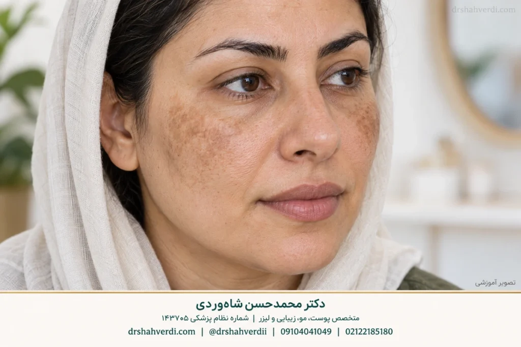
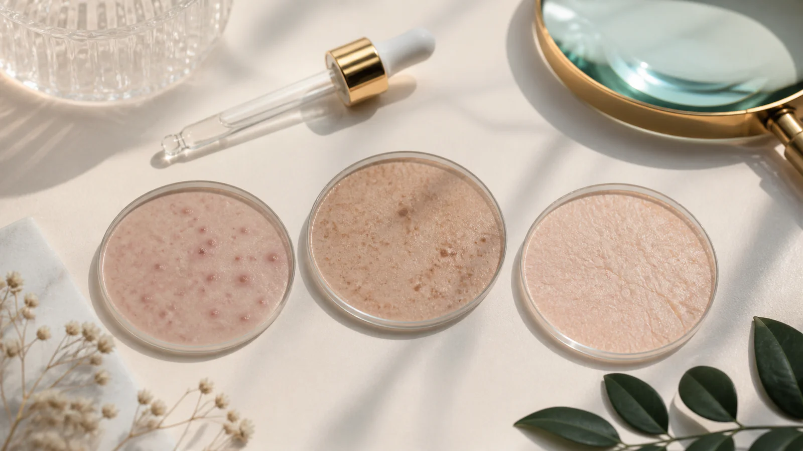
Hyperpigmentation means an area of skin has more colour than the surrounding skin; the term does not identify the cause. Melasma, marks left after inflammation and sunspots, also called solar lentigines, can look similar in a photograph while needing different care. This guide helps you describe the pattern and prepare for assessment. It cannot turn a comparison table into a diagnosis.
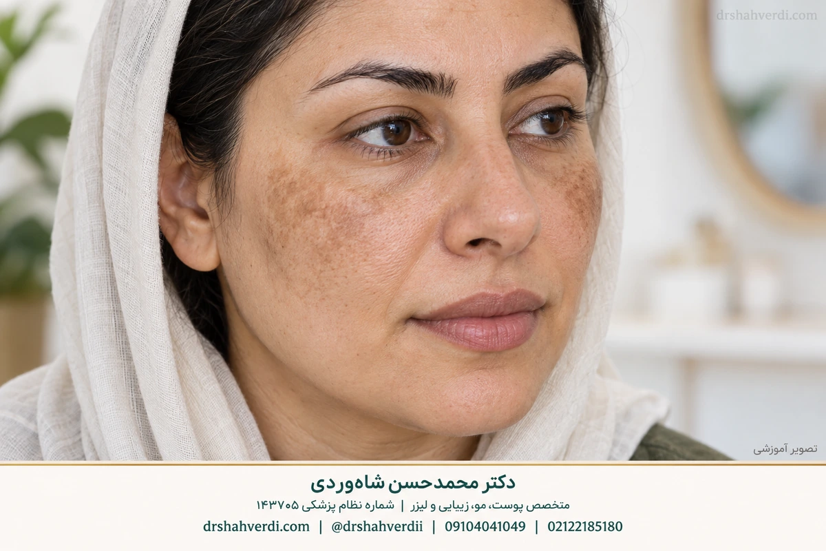
Why the cause matters
A dark mark is a description, not a treatment instruction. Note when it appeared, whether another skin problem came first and what has changed since then. A flat mark and an indented acne scar are different problems. Shadows under the eyes can also have causes beyond pigmentation. Use these distinctions to explain your concern rather than choose a cream solely from its label.
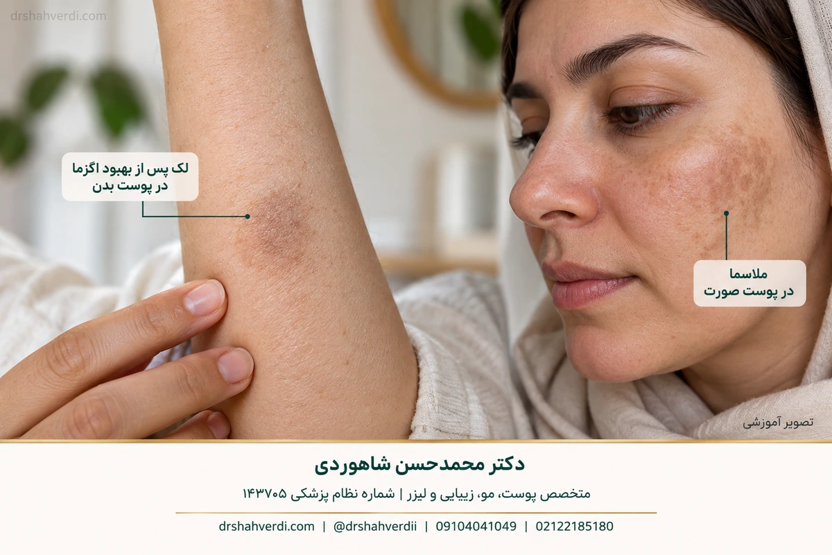
What is acquired dermal macular hyperpigmentation?
Acquired dermal macular hyperpigmentation is a term used for a group of acquired grey-brown pigmentation disorders. It should not be treated as another name for melasma or assumed to need the same treatment. Clinical assessment considers the distribution, symptoms, previous inflammation and other findings. Our lichen planus pigmentosus guide explains one relevant differential diagnosis.
Melasma is more common in women but occurs in men too. Chloasma is another name, frequently used in pregnancy discussions; the English melasma guide explains the terminology. Hyperpigmentation is a colour change, whereas rosacea commonly involves redness and inflammation and vitiligo involves loss of pigment. More than one condition can coexist. A name or photograph cannot resolve that distinction for an individual.
Hyperpigmentation means increased colour, while hypopigmentation means reduced colour. These terms describe an appearance and do not identify its cause. Vitiligo is one diagnosis involving pigment loss; every lighter patch should not be labelled vitiligo.
Compare three common patterns
Scroll sideways to see all columns.
| Pattern | Typical clue | Limit of the clue |
|---|---|---|
| Melasma | Broader facial patches, often on both sides | Symmetry does not prove melasma |
| PIH (post-inflammatory hyperpigmentation) | Marks where inflammation or injury occurred | Inflammation may still be active |
| Sunspots (solar lentigines) | Persistent, more discrete spots on sun-exposed skin | A new spot still needs assessment |
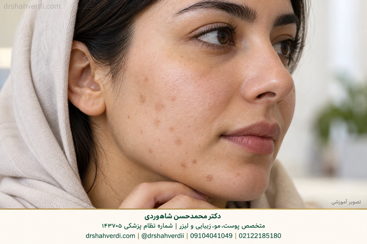
Read the focused guides to melasma, PIH and solar lentigines.
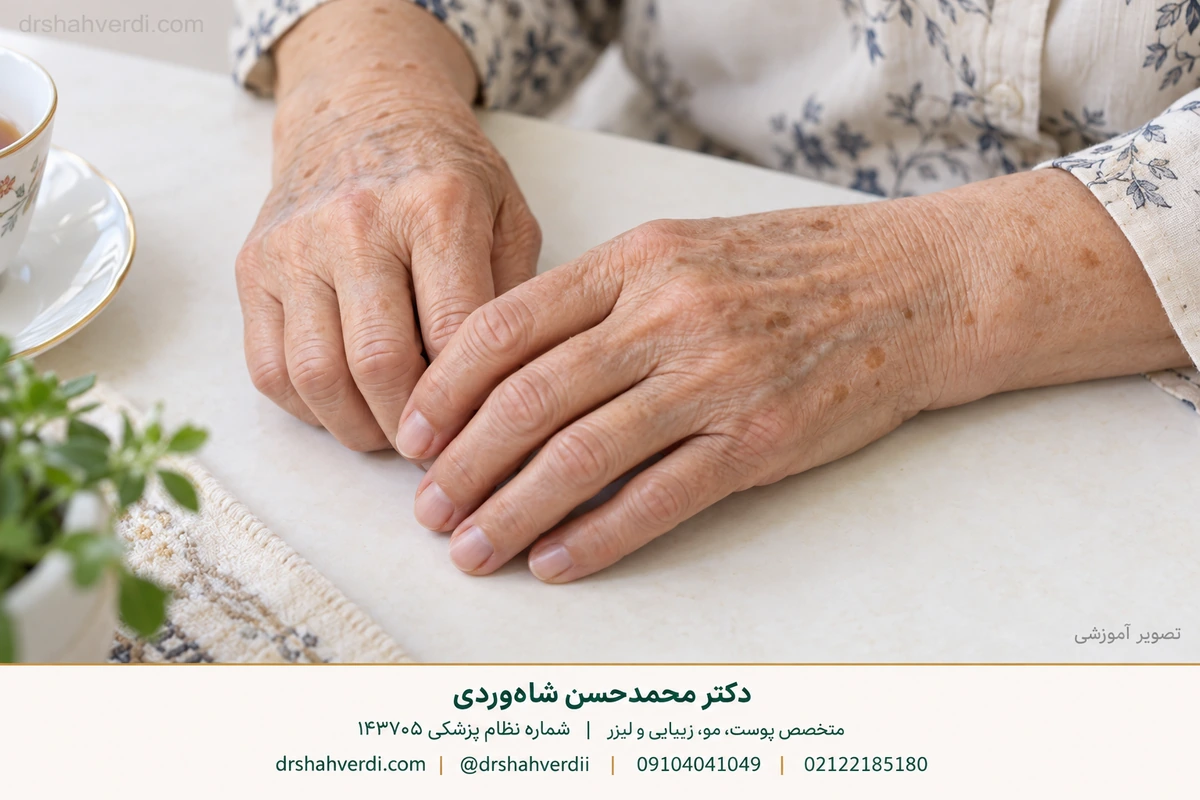
What does an ICD code tell you about dark spots?
Hyperpigmentation and dark spots describe an appearance; they do not provide one sufficiently specific diagnosis or one universal ICD code. PIH, melasma and solar lentigines are different conditions. A code should follow the clinician’s documented diagnosis and the classification version used by the relevant healthcare system. WHO explains the purpose of ICD.
ICD-10 and the US clinical modification, ICD-10-CM, should not be assumed interchangeable for every question or country. CDC describes ICD-10-CM as the system used for medical diagnoses in the United States. A coding search does not establish your diagnosis, treatment or insurance coverage. Ask the clinician or coding team to clarify which system applies to your record.
Other patterns that can resemble a dark spot
Scroll sideways to see all columns.
| Condition or group | Why the history changes the next step |
|---|---|
| Exogenous ochronosis | Blue-black pigmentation can develop with prolonged use of certain lightening products; it must be distinguished from treatment-resistant melasma. |
| Lichen planus pigmentosus | One of the acquired grey-brown pigment conditions that may overlap in appearance. Its full explanation remains in the existing LPP guide (Persian). |
| Ashy dermatosis (EDP) | Grey patches can affect the face, neck or trunk; some have an early reddish edge. That edge is not always present, and related disorders overlap. DermNet EDP. |
| Pigmented contact dermatitis | Gradual grey-brown facial darkening makes cosmetic, fragrance and hair-dye history relevant. A clinician may use patch testing to investigate a contact allergy; colour alone cannot identify the culprit. Original patch-test study. |
| Hori naevus | Acquired brown-grey or blue-grey dermal pigmentation typically affects both sides of the face. It is not established as melasma by its colour. DermNet Ota/Hori. |
| Naevus of Ota | Forehead or eye-area pigmentation may be present from childhood or become apparent later. The eye itself can be involved; eye assessment is needed in that situation. DermNet Ota/Hori. |
| Pityriasis versicolor or confluent and reticulated papillomatosis | Scaling, texture and a trunk/neck pattern may suggest a different disorder; a facial anti-pigment prescription should not be copied onto it. |
Acquired dermal macular hyperpigmentation is an umbrella term used for related acquired pigment disorders, including LPP and ashy dermatosis. It does not mean all grey-brown patches have one cause. The existing LPP guide (Persian) owns its full explanation. DermNet ADMH and PCDS facial/neck differential guidance provide original clinical context.
Lentigo is singular and lentigines is plural; solar lentigo is a specific sun-related type, rather than a name for every lentigo. Merriam-Webster Medical gives lentigines as len-ˈtij-ə-ˌnēz. Its solar entry provides the other word’s pronunciation. Pronunciation does not determine whether a particular spot is benign.
Pityriasis versicolor is a yeast-associated condition with finely scaly patches that can be darker, lighter or pink, often on the trunk or neck. It needs its own assessment and, when confirmed, antifungal treatment rather than a melasma prescription. Confluent and reticulated papillomatosis can cause brown, scaly or raised patches with a net-like edge, often on the upper trunk or neck. It can resemble versicolor or acanthosis nigricans and cannot be diagnosed by colour alone; it should not be assumed to be a yeast infection or proof of diabetes.
Use the right official code lookup
For a US ICD-10-CM record, open the CDC official browser, choose the fiscal year relevant to the service date, search the clinician’s recorded diagnosis, and read the tabular notes and exclusions. Generic dark spots do not have one universal code. Another country may use a different classification or modification; confirm the system with the clinician or coding team. These steps support a lookup and do not assign a personal diagnosis or insurance decision.
The history behind the colour
Post-inflammatory hyperpigmentation, or PIH, follows processes such as acne, eczema, irritation or injury. It can persist after the original problem improves, and it may be more noticeable or lasting in darker skin. Show the dermatologist where the original rash or spots occurred. A colour seen in a photograph cannot reliably identify depth or distinguish every possible diagnosis. DermNet PIH.
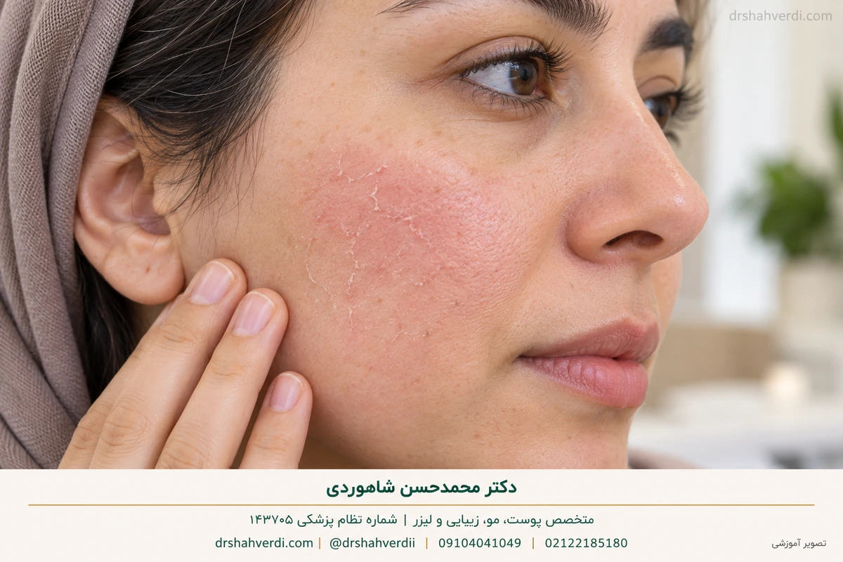
When location or general darkening needs medical review
Record whether a patch followed acne, eczema, hair removal, a medicine or a procedure. Pigment on the neck, around the mouth or over a wider body area can have a different explanation from melasma. Thick or velvety fold changes should be assessed; the acanthosis nigricans guide (Persian) is a focused route. A dark patch alone does not diagnose diabetes, liver disease or an adrenal disorder.
If more general darkening is new and accompanied by fatigue, weight loss, dizziness or other systemic symptoms, arrange medical assessment rather than a cosmetic procedure. Mouth or gum pigmentation also needs its own history and examination. Severe weakness, fainting or being acutely unwell needs prompt medical care. The appropriate tests depend on the findings; this article does not supply a routine test panel for all dark spots. For detailed location-related care, use the existing body-pigmentation guide (Persian).
PIH is usually assessed from the preceding inflammation and skin pattern. Histology means examining an actual tissue specimen when one is clinically needed; pigment in a specimen must be interpreted alongside the history and examination. A pathology term or online photograph does not assign a diagnosis to you. DermNet PIH provides original clinical context.
Dark, thick, velvety skin may be acanthosis nigricans; sometimes it is associated with insulin resistance or prediabetes, but appearance alone does not diagnose diabetes. New widespread darkening, including the mouth or old scars, together with persistent fatigue, weakness, weight loss or dizziness warrants medical assessment. Addison’s disease is one possible cause in this wider context; an isolated facial patch is not diagnostic. “Liver spots” commonly describes certain age-related sunspots and does not diagnose liver disease; it is not a reason for a detox remedy or automatic liver tests.
Matching care to the problem
When acne or eczema continues, controlling that condition is part of preventing further marks. Selected topical treatment may help some pigmentation. Discrete benign sunspots have a different treatment discussion from melasma, and a device recommendation should follow diagnosis. Neither the most expensive product nor the strongest procedure ensures the best outcome. Review our treatment-options guide for practical questions.

Match the first step to the cause
Scroll sideways to see all columns.
| Pattern after assessment | Usual decision focus |
|---|---|
| Melasma | Light protection, a tolerable selected medicine and a maintenance plan; return of pigment remains possible. |
| PIH | Control active acne, eczema or irritation and prevent new injury. Flat colour is different from a depressed or raised scar. |
| Confirmed solar lentigo | Decide whether cosmetic treatment is wanted after confirming the lesion. A new or changing “sunspot” must be examined before removal. |
| Another pigment disorder | Use diagnosis-specific care rather than escalating a melasma cream. |
Natural skin colour is not a diagnosis. Irritating whitening recipes can create additional PIH in any skin tone; gentle care matters particularly where pigment changes are more persistent. A new, bleeding, non-healing or changing spot should go to examination, including the broader sun-related skin-lesion guide (Persian) where relevant.
Which spots deserve earlier assessment?
Arrange assessment for a lesion that is new and changing, unusually different from the others, bleeding or failing to heal. Do not assume age spots are harmless simply because they occur on sun-exposed skin. A suspicious lesion should be examined before laser or another cosmetic attempt to remove its colour. Avoid using online comparisons to decide that biopsy or follow-up is unnecessary. Solar-lentigo assessment.
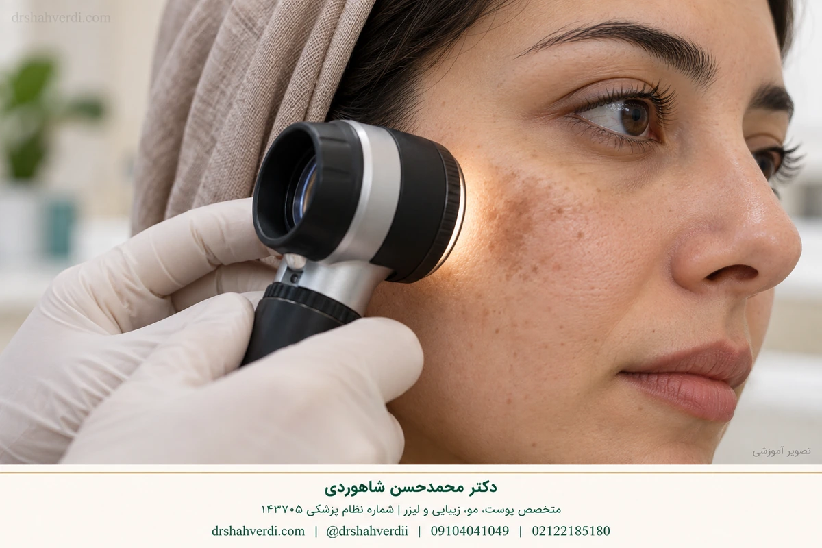
A confirmed solar lentigo is a benign diagnosis, but melanoma can sometimes resemble a dark spot. This comparison cannot exclude skin cancer. A new, changing, bleeding or non-healing lesion needs examination before a cosmetic removal or lightening treatment. AAD explains why an apparent age spot should be checked before treatment.
Reduce irritation and further darkening
Keep the routine gentle and protect exposed skin from sunlight. Avoid picking acne and using lemon juice, abrasive scrubs or repeated home peels. A product that irritates the skin can create additional dark marks. Check unfamiliar brightening products for a clear ingredient list rather than trusting unqualified promises. If a product repeatedly burns or stings, seek guidance about changing the routine. AAD dark-spots advice.
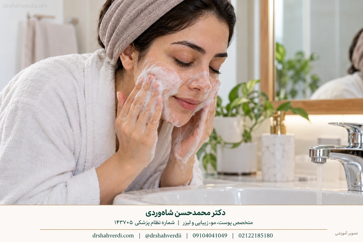
Choose a broad-spectrum sunscreen with SPF 30 or higher, together with shade and a broad-brimmed hat. Visible light can worsen melasma, especially in darker skin tones; a tinted formula containing iron oxides adds relevant protection. A mineral label, zinc oxide alone or a cosmetic shade does not confirm that a product protects adequately against visible light. Reapply outdoors according to the label, usually every two hours and after sweating or washing. Choose a tolerable texture you can use consistently; an advertising claim such as “best” does not replace label reading. AAD self-care guidance and our focused sunscreen guide (Persian) explain the distinction.
Prepare a clear record
Bring previous photographs, medicine names, treatment dates and the sequence of any irritation. Explain whether the pigmentation appeared after a procedure and whether pain, itch or redness remains. Several diagnoses can coexist, so one face may need more than one approach. For assessment with Dr Mohammadhasan Shahverdi, see the Tehran consultation guide.

Describe the sequence: what appeared first, what treatment followed and what changed afterwards. That history often helps more than a colour label alone.
Questions about dark marks
Are freckles the same as age spots?
Freckles often darken with sunlight and fade seasonally. Solar lentigines usually persist. Individual spots still need assessment when the diagnosis is uncertain.
Is a brown acne mark an acne scar?
PIH is a colour change; a scar changes the skin’s structure. They can coexist, but their treatment goals differ.
Can a laser cause hyperpigmentation?
Yes. Procedure-related inflammation can leave pigmentation. Explain the treatment and timing to your clinician before starting another procedure.
Does PCOS explain dark marks around my mouth, chin or face?
A PCOS diagnosis does not identify the cause of every dark mark. A flat mark following acne, a rash, hair removal or an irritating product can have a different explanation from a broader melasma patch. Note what appeared first and whether there was redness, itch or inflammation. Darker colour alone is not a diagnostic symptom that confirms PCOS or insulin resistance. The patterns above help you describe the change; an examination determines which explanation fits. AAD explains common causes of dark marks.
Is there a specific cream for pigmentation when I have PCOS?
There is no single cream chosen from the ovarian diagnosis alone. Ask whether the target is inflammation, a mark left by it, melasma or another condition. The care options above explain why active acne or irritation needs attention alongside the colour. Tell the clinician about pregnancy or breastfeeding plans and previous reactions before choosing a product. Do not copy a facial lightening treatment onto dark, thick or velvety folds; that pattern belongs in the acanthosis assessment section. Metabolic care, when indicated, does not guarantee that every pigment mark will disappear. AAD explains the variable response of acanthosis to treatment.
Can a Reddit success story tell me which treatment will work?
No. People may use “hyperpigmentation” for different diagnoses, and a post does not reliably establish the original cause, all medicines used or whether the result lasted. Bring the proposed product name as a question for your visit rather than copy the regimen. The useful comparison is with your diagnosis and treatment history, not another person’s photograph. The assessment section explains what to record.
Scientific sources
- DermNet: post-inflammatory hyperpigmentation
- DermNet: solar lentigo
- American Academy of Dermatology: How to fade dark spots
- American Academy of Dermatology: Melasma signs and symptoms
- DermNet: ADMH
- WHO: International Classification of Diseases
- CDC: ICD-10-CM
- PCDS: face and neck hyperpigmentation
- Merriam-Webster Medical: lentigines pronunciation
- Merriam-Webster: solar pronunciation
- DermNet: pityriasis versicolor
- DermNet: confluent and reticulated papillomatosis
- CDC official ICD-10-CM browser
- AAD: acanthosis nigricans
- NIDDK: adrenal insufficiency and Addison disease symptoms
- AAD: examine apparent age spots before treatment
- AAD: melasma self-care
بعد از این مطلب، چه بخوانیم؟
ذخیرهسازی اختیاری است و فقط در همین مرورگر انجام میشود؛ بدون حساب کاربری و بدون ارسال فهرست به سایت. روی دستگاه مشترک، دیگران هم میتوانند فهرست را ببینند. با پاکشدن دادههای مرورگر، فهرست از بین میرود.
مطالب ذخیرهشدهٔ من
هنوز مطلبی ذخیره نکردهاید. کنار راهنماهای پیشنهادی، «برای بعد نگه دار» را انتخاب کنید.
همهٔ مطالب ذخیرهشده از این مرورگر حذف شوند؟
حالا دوست دارید چه چیزی روشنتر شود؟
انتخاب شما فقط ترتیب پیشنهادها را عوض میکند؛ برنامهٔ درمانی نیست.


نظر شما، ادامهٔ این مطلب است
کدام بخش برایتان مفید بود؟ چه پرسشی دربارهٔ موضوع مقاله باقی مانده است؟
دیدگاهها پس از بررسی منتشر میشوند. این بخش عمومی است؛ لطفاً شماره تماس، تصویر، نتیجه آزمایش یا اطلاعات شناسایی و پزشکی خصوصی خود را ننویسید. دیدگاهها جایگزین معاینه و مشاوره فردی نیستند.
ثبت دیدگاه برای این مطلب در حال حاضر بسته است.